Dog Skin Tag on Nipple: Symptoms, Diagnosis, and Vet Treatment Guide
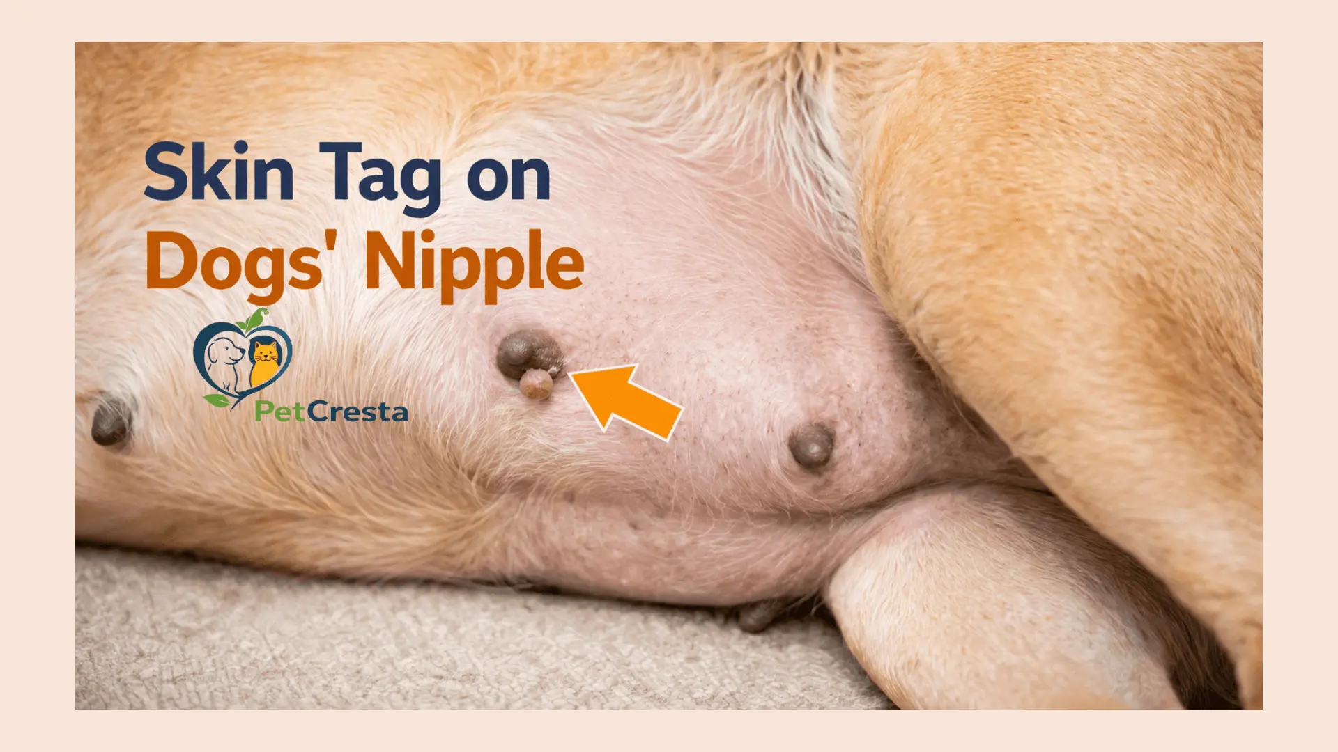
A small growth on a dog’s nipple is often called a skin tags on dogs nipple by owners. Sometimes that description fits. Sometimes it does not.
This body site needs more caution than an ordinary skin bump. The nipple sits within the mammary chain, so a visible growth may involve the skin, the nipple itself, or nearby mammary tissue.
The first job is to check a few basics. Look at the feel, the pattern of change, the surface, and whether there is pain, discharge, redness, or licking. A matching nipple on the other side also matters, because a normal structure can sometimes be mistaken for a new lump.
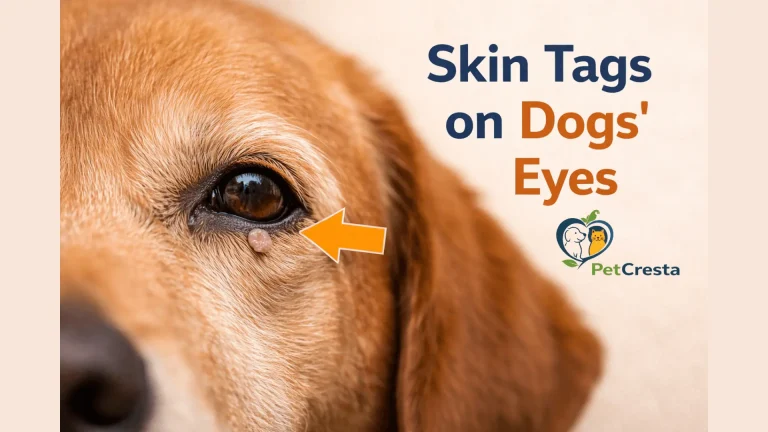
Do not cut it, tie it off, squeeze it, or apply human skin products. If a similar bump is closer to the eyelid area, skin tags on a dog’s eyes may be the better internal reading path.
Quick Verdict: Is This Nipple Area Growth Likely Minor Or Worth Checking Soon?
☐ A soft, stable, surface-level flap is usually less concerning than a firm or fixed lump.
☐ A bump that is growing, bleeding, painful, ulcerated, swollen, or producing discharge needs a faster veterinary check.
☐ Because this growth is on or near a nipple, home assumptions are less reliable than they would be for a simple skin bump elsewhere.
Dog Nipple Growth At A Glance
This table helps with triage, not diagnosis. A small lesion can still need testing if the location or feel is concerning.
| Feature | Lower concern pattern | More concerning pattern |
|---|---|---|
| Feel | Soft, flexible, surface-level | Firm, nodular, deeper, fixed |
| Shape | Small flap or stalk | Round lump, thickened area, irregular mass |
| Change | Stable | Enlarging or changing |
| Surface | Smooth and dry | Crusty, bleeding, ulcerated, oozing |
| Dog’s response | Ignores it | Licking, chewing, pain on touch |
| Discharge | None | Bloody, cloudy, or pus-like |
| Pattern | Matching structure on both sides | New isolated bump or uneven change |
| Next step | Monitor until the exam | Book sooner, urgent if severe signs are present |
What Is Skin Tag On Dogs Nipple
Owners often use this phrase for any fleshy bump in the nipple area. That is understandable, but the same label may be used for different problems.
In many cases, the phrase means a small flap, bump, or hanging bit of tissue on or beside the nipple. The challenge is that the visible part may not tell the full story. A lesion may arise from the skin itself, from the nipple, or from tissue just under it.
That is why this location deserves more care than a random belly bump. A growth on skin away from the nipples is one thing. A growth attached to the nipple line is another, because mammary tissue may be part of the picture.
Types: Could This Be a Normal Nipple, a Wart, or a Mammary Lump?
A nipple area bump should be compared with common look-alikes before anyone assumes it is a harmless tag.
| Possibility | How it tends to look | How it tends to feel | What raises concern | Typical next step |
|---|---|---|---|---|
| Normal nipple | Smooth, even, placed in line with the other nipples | Soft to slightly firm | Sudden size change, redness, discharge | Compare with the matching side |
| Skin level tag | Small flap or narrow stalk on the surface | Soft, movable with the skin | Growth, rubbing, bleeding, repeated irritation | Mention at the next visit or sooner if changing |
| Wart-like growth | Rougher or cauliflower-like surface | Often surface-level | Fast growth, ulceration, multiple lesions | Vet exam to identify the type |
| Mammary lump | Rounder or thicker area under or beside the nipple | Firmer, more rooted, less movable | Ongoing growth, pain, discharge, fixation | Prompt veterinary exam |
| Inflamed gland | Swollen, warm, red, sometimes enlarged | Tender or painful | Heat, discharge, illness, nursing history | Quick exam, same day if severe |
A normal nipple can be mistaken for a skin growth, especially in dogs with small or flat mammary tissue. A wart can also look tag-like at first. The more a lesion feels deep, thick, attached, or newly changing, the less useful the word “skin tag” becomes.
What It May Look And Feel Like
Some nipple area growths stay soft and surface-level. Others feel firm, thick, or more attached, which makes them more concerned. For bumps higher on the muzzle or outer facial skin, see skin tag on a dog’s face.
Features That Fit A Simple Skin Level Growth
A more reassuring lesion is often small, soft, and superficial. It may sit on a narrow stalk and move with the skin when touched gently.
It may stay the same size for a long time and may not bother the dog unless it is rubbed, scratched, or chewed.
Features That Need More Caution
A lesion deserves more concern if it feels firm, thick, nodular, or fixed in place. The same is true if it looks deeper than the surface or seems attached within tissue rather than sitting on top of it.
Bleeding, crusting, swelling, warmth, discharge, pain, or steady enlargement make the area less reassuring.
Quick Comparison Box
| Clue | Surface growth | Deeper nipple area mass |
| Depth | On the skin | Within or under tissue |
| Texture | Soft, flexible | Firm, dense, nodular |
| Mobility | Moves with skin | Feels more attached |
| Irritation | Mild if rubbed | May be painful or inflamed |
| Change | Often slow or stable | More likely to enlarge |
When To Watch, When To Book, And When It Is More Urgent
A stable bump can sometimes wait a little. A growing, painful, bleeding, or discharging lesion should be checked sooner. If the bump is lower on the limb or close to the toes, a skin tag on a dog’s paw is the better internal match.
Lower Concern Pattern
A bump is less worrying when it is very small, soft, stable, and not causing trouble. The surrounding skin stays normal. The dog is not licking it. There is no discharge, swelling, or pain.
Even then, this location still belongs on the list for a veterinary check because nipple area growths can overlap in appearance.
Book A Vet Visit Soon
Move the visit up if the lump is getting bigger, feels firm, has more than one nearby nodule, or starts to look crusted or irritated.
A new growth in the mammary region should not be watched casually for too long, even if the surface still looks mild.
Same Day Or Urgent Care Signs
Seek quicker care if the area becomes very swollen, hot, dark, severely painful, or starts producing bloody or pus-like discharge.
A dog that seems feverish, dull, weak, or unwell along with a nipple area problem, needs faster attention. This matters even more in a dog that is nursing or has recently weaned.
Causes: Why A Growth Can Show Up On Or Near A Nipple
Not every dog skin tag on nipple comes from the same source. When the lesion sits over a pressure point rather than the soft part of the limb, a skin tag on a dog’s elbow is a better internal topic.
Skin Level Growth On The Surface
Some lesions arise from the skin itself and behave like a superficial growth. These are usually softer and more surface-level than a deeper mass.
Nipple Associated Lesion
Some bumps sit directly on the nipple tissue. They may still look small from the outside, but the exact attachment site matters because the nipple is not just ordinary skin.
Mammary Tissue Change
Some lesions involve tissue beneath or around the nipple. These may feel firmer, more rooted, or less mobile than a simple surface growth.
Pregnancy, Nursing, And Recent Weaning
A nursing or recently weaned dog needs extra care in this area. Swelling, heat, redness, pain, or discharge may point more toward mammary inflammation than a simple skin lesion.
That context matters because a sore mammary gland can be mistaken for a small growth at first glance.
Risk Factors And Context That Matter In This Location
Age, sex, and reproductive history help shape how a nipple area lump is interpreted, even though they do not identify the lesion by themselves.
Female Dogs
Most mammary problems occur in females. That does not mean every bump is serious, but a nipple area lesion in a female dog deserves a careful exam rather than a guess.
Male Dogs
Male dogs have nipples and can still develop lesions in this area. Mammary tumors are much less common in males, but a new lump still needs proper assessment.
Older Dogs
Older dogs often develop more lumps over time. Some are benign surface growths. Some are not. Age alone does not define the lesion, but it makes the new nipple area change more important to evaluate.
Hormone And Reproductive Status
Intact or later spayed females have greater mammary relevance than dogs spayed early in life. Pregnancy, lactation, and recent weaning also change how swelling or nipple area thickening should be interpreted.
What To Track At Home Before The Appointment
A few simple details can make the visit more useful. Tracking size, feel, discharge, and change over time helps show whether the lesion seems stable or is becoming more concerning.
What To Note
Write down which nipple is involved and when the bump was first noticed. Note whether it feels soft or firm, surface level or deeper.
Also note any growth, discharge, crusting, bleeding, licking, swelling, or pain on touch. Check whether there is a matching structure on the other side.
How To Document It
Take a clear photo in good light and repeat from the same angle if the lesion changes. A ruler or coin nearby can help show size.
Also note whether the dog is intact, pregnant, nursing, or recently weaned.
What Not To Do While Waiting
Do not trim the lesion. Do not tie it off. Do not squeeze it. Do not apply human wart or skin products.
Those steps can irritate the area and make the assessment harder.
Home Tracking Checklist
☐ Which nipple is involved
☐ Date first noticed
☐ Soft or firm
☐ Surface level or deeper
☐ Stable or growing
☐ Any discharge
☐ Any bleeding or crusting
☐ Licking or pain
☐ Matching structure on the other side
☐ Pregnant, nursing, or recently weaned
☐ Photo taken
What the Vet May Do to Diagnose It
A nipple area lesion is diagnosed by location, feel, and testing when needed.
Physical Exam And Mammary Chain Palpation
The vet checks whether the lesion seems limited to the skin or connected to deeper tissue. The rest of the mammary chain may also be examined because more than one lump can occur.
Nearby lymph nodes may be checked depending on the findings.
Fine Needle Aspiration, Biopsy, And Histopathology
A fine needle aspiration may be used to collect cells. This can help distinguish some surface lesions from mammary-associated disease, but it does not always give the full answer.
If the lesion is suspicious or unclear, biopsy or removal followed by histopathology gives a more reliable diagnosis.
Imaging And Staging When Needed
If mammary involvement is suspected, some dogs need more testing. This may include chest imaging, abdominal imaging, bloodwork, or lymph node assessment.
Not every lesion needs all of these steps. The plan depends on what the lump feels like and how concerning it appears.
Why Earlier Evaluation Helps
Earlier evaluation helps because a smaller, quieter lesion is easier to assess and plan for. Waiting until the area grows, ulcerates, or becomes inflamed can make the picture more complicated.
Treatment And Removal Of Skin Tag On Dogs Nipple
A dog nipple skin tag treatment depends on whether the lesion is superficial or tied to deeper nipple area tissue. The priority is correct diagnosis and safe removal when needed.
When Monitoring May Be Reasonable
Monitoring may be reasonable when the lesion is very small, soft, stable, clearly superficial, and not irritated. That choice should follow a veterinary exam, not home guesswork.
A monitored lesion still needs size checks and follow-up if anything changes.
When Testing Or Removal Is More Likely
Removal or further testing is more likely when the lesion is growing, bleeding, painful, ulcerated, producing discharge, or difficult to identify confidently on exam.
Firmness, deeper attachment, and concern for mammary involvement also make removal or tissue sampling more likely.
What Dog Skin Tag On Nipple Treatment May Involve
A true superficial lesion on or beside the nipple may be removed as a local procedure if it is being traumatized or if the diagnosis is uncertain.
If the lesion appears tied to the nipple or nearby mammary tissue, treatment may involve a more careful surgical plan based on the exam and test results. The goal is not cosmetic removal. The goal is accurate diagnosis and complete management of the lesion in that location.
What Dog Skin Tag Nipple Surgery May Involve
Surgery may range from the removal of a small surface lesion to the excision of a deeper nipple area mass. The exact plan depends on depth, tissue involvement, imaging findings, and how suspicious the lesion appears before surgery.
Tissue is often submitted for microscopic review after removal, so the next steps can be planned correctly.
Recovery After Removal
Recovery depends on the size and depth of the procedure, but most dogs need close incision monitoring and strict control of licking.
Recovery checklist:
☐ Use the cone as directed
☐ Keep the incision clean and dry
☐ Restrict rubbing, licking, and rough activity
☐ Return for recheck and pathology review if tissue was submitted
Dog Skin Tag On Nipple Cost: What Can Affect Price
The cost of a dog nipple skin tag depends on the workup as much as the removal itself. A simple exam costs less than a visit that also includes cell sampling, biopsy, imaging, anesthesia, surgery, and laboratory review.
A small surface lesion removed during another planned anesthetic event may cost far less than a deeper mammary region surgery. That is why nipple area growths should not be priced like a routine cosmetic skin procedure.
Dog Skin Tag On Nipple Home Removal Is Not Safe
Home removal is not safe for nipple area lesions. Even a small bump can bleed heavily, become infected, or turn out to involve deeper tissue.
This location needs a diagnosis before treatment. Cutting, tying off, or using human removers can delay proper care and make later assessment harder.
Conclusion
A dog skin tag nipple may be a simple surface lesion, but this is not a location where appearance alone should be trusted too much.
The safest approach is to judge the lesion by its location, feel, change over time, and whether there is pain, discharge, swelling, or deeper attachment. A small stable bump still deserves mention at the next visit. A firm, changing, bleeding, or painful one deserves a prompt exam.
FAQ’s About Skin Tag on Dog Nipples
Yes. A normal nipple or a matching structure on the other side can sometimes be mistaken for a new lump, especially in dogs with small mammary tissue or uneven hair cover. That is one reason location and symmetry matter. If one side suddenly looks different, feels thicker, or becomes irritated, it should be checked rather than assumed normal.
Sometimes yes. A wart-like lesion may have a rougher or more irregular surface, while a skin tag is often softer and smoother. In real patients, though, these can overlap in appearance, and neither look can rule out a deeper problem near the nipple. A new or changing bump still needs proper context and, in some cases, testing.
Yes. Some mammary region lumps start small and do not look dramatic on the surface. That is why a quiet appearance does not always mean a minor problem. If a bump feels firm, rooted, or continues to enlarge over time, a veterinary assessment should move up.
Any new lump in this region deserves a veterinary look, even if it seems mild at first. The need becomes more urgent if the lesion is growing, firm, painful, swollen, bleeding, crusted, or producing discharge. Faster care also matters if the dog seems unwell or is nursing.
Yes. Male dogs have nipples and can develop growths in that area. Mammary tumors are much less common in males than in females, but a new lump still needs the same careful exam because surface lesions and deeper masses can look similar early on.
A more concerning lesion often feels firmer, thicker, or more attached than a simple surface flap. It may seem rooted in the tissue instead of moving freely with the skin. That does not diagnose the lump by touch alone, but it does help explain why some masses need a faster workup.
Yes. Bleeding can happen when a lesion is rubbed, scratched, or ulcerated. Discharge is more concerning because it may reflect inflammation, mammary disease, infection, or a more irritated lesion. Bloody, cloudy, or pus-like material should not be ignored.
There is no single price that fits every case. Cost changes with the exam, whether testing is needed first, how deep the lesion is, whether anesthesia is required, and whether tissue is sent for pathology. A small superficial removal is very different from surgery on a suspicious mammary region mass.
Most dogs need incision checks, activity control, and prevention of licking while the site heals. A cone is often needed, and follow-up matters if tissue was submitted for microscopic review. Recovery is usually simpler for a small surface excision than for a deeper nipple or mammary surgery.
Yes. Some nipple area lesions are benign and remain superficial for a long time. The challenge is that harmless surface growths and more important mammary region lesions can overlap in appearance. That is why a calm, location-specific exam is still the safest next step.
References
- VCA Animal Hospitals, Benign Mammary Tumors in Dogs
- VCA Animal Hospitals, Malignant Mammary Tumors in Dogs
- VCA Animal Hospitals, Mastitis in Dogs
- Cornell Riney Canine Health Center, Mammary Cancer
- Merck Veterinary Manual, Mammary Breast Tumors in Dogs
- PDSA, Skin Lumps and Tumours in Dogs
- PDSA, Mammary Problems in Dogs: An Overview
- PetMD, Skin Tags on Dogs