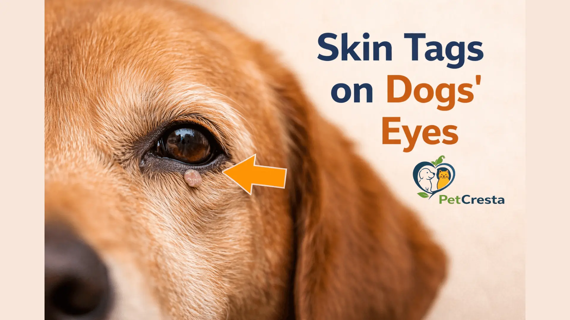
Dog Skin Tag on Eye: Symptoms, Diagnosis, and Vet Treatment Guide
A small bump near a dog’s eye can look minor at first. This area deserves more attention than many other skin growth sites because even a harmless-looking lesion can irritate the eye surface, affect blinking, or become harder to manage if it keeps growing.
In many cases, this points to a bump on the eyelid or the skin around the eye, not a growth on the eyeball itself. Many eyelid masses in dogs are benign, but the location still matters because comfort, tear spread, and corneal protection come first.
Quick Answer: What Most Dog Owners Need to Know First
- Many bumps, called a skin tag, near a dog’s eye are on the eyelid or nearby skin, not on the eyeball.
- Many eyelid lumps are benign, but this location matters because blinking can make even a small bump irritating.
- Redness, tearing, discharge, squinting, rubbing, bleeding, crusting, or clear contact with the eye requires a prompt vet visit.
- Home removal is not safe in this area.
Skin Tags on Dogs Eye: Characteristics At A Glance
| Feature | Common pattern |
| Usual location | Eyelid margin, upper lid, lower lid, or skin beside the eye |
| Common appearance | Small raised bump, soft flap, smooth nodule, or lobulated growth |
| Common color | Pink, tan, brown, gray, or darkly pigmented color |
| Common age group | More often seen in middle-aged and older dogs |
| Symptoms that matter | Tearing, squinting, redness, discharge, rubbing, blinking, discomfort |
| What raises concern | Growth over time, bleeding, crusting, corneal contact, trouble closing the lid |
| What owners should do next | Monitor only after a vet exam, or book a visit sooner if irritation is present |
What Is a Dog Eyelid Skin Tag
A dog skin tag on eye usually means a small flap, bump, or raised lesion on the eyelid or nearby skin. Owners may use this phrase for a growth on the lid margin, above the eye, below the eye, or close to the inner corner. A true growth on the eyeball itself is different and needs faster veterinary assessment.
This area matters because the eyelid moves across the eye with every blink. A tiny lesion on the lid edge may be more irritating than a slightly larger one on nearby skin. Position often matters as much as size.
What These Bumps Can Be
A bump in this area may look like a skin tag at home, but several eyelid conditions can look similar. Some are simple and slow-growing. Others need treatment because they irritate the cornea, distort the eyelid, or make the diagnosis less clear.
The main point is that skin tag is often a description, not a diagnosis. Around the eye, the label matters less than how the bump behaves and whether it affects comfort.
Skin Tag On Dog Eye Causes
A small eyelid growth may develop from normal skin tissue, blocked oil glands in the eyelid, age-related change, mild chronic irritation, or another benign eyelid mass. Some lesions appear slowly over time. Others become noticeable only when they start rubbing the eye or catching debris.
Inflammation can also create a lump-like appearance. A blocked gland may swell. A stye-like lesion may look red and irritated. Pigmented growths can appear darker and often worry owners more, even when they are not always aggressive.
Types Of Dog Eye Growths
| Growth type | What it may look like | Why it matters |
|---|---|---|
| Skin tag like growth | Soft flap or small flesh colored bump | May stay minor if away from the cornea |
| Meibomian gland growth | Smooth or lobulated bump on lid margin | Common in older dogs and often irritates the blinking |
| Papilloma | Wart-like or rough-surfaced lesion | Can resemble a skin tag at home |
| Chalazion | Swollen blocked gland | May cause local irritation or lid swelling |
| Hordeolum | Tender inflamed bump | More likely to look red or sore |
| Pigmented mass | Dark bump or plaque | Needs closer evaluation if changing or irregular |
| Third eyelid issue | Pink or fleshy tissue near the inner eye corner | Often mistaken for a skin tag by owners |
If the growth is not limited to the eyelid area, a skin tag on dogs face may help explain nearby facial bumps in more detail.
Common Skin Tag-Like Eyelid Growths
A meibomian gland adenoma is one of the more common benign eyelid growths in dogs. It develops from an oil gland in the eyelid and may appear as a rounded or slightly uneven bump along the lid edge. Papillomas can also occur in this region and may look rougher or more wart-like.
Some lesions are pale and soft. Others are darker and firmer. Appearance can point in a direction, but appearance alone cannot confirm what the bump is.
Skin Tag on Dog Eye Vs Tumor Dog Eye
A skin tag near a dog’s eye is usually described as a small, soft, flesh colored growth that may hang slightly from the eyelid or nearby skin. A tumor is a broader term for an abnormal growth. Some eyelid tumors are benign, while others are more concerning.
The word tumor does not automatically mean cancer. Around the eye, the bigger issue is often whether the lesion is rubbing the cornea, changing lid shape, or becoming inflamed or ulcerated.
| Feature | Skin tag like growth | More concerning mass |
|---|---|---|
| Texture | Soft and flexible | Firm, thick, fixed, or irregular |
| Shape | Small flap or smooth bump | Nodular, uneven, broad-based |
| Growth pattern | Often slow and stable | May enlarge, change shape, or recur after irritation |
| Surface | Usually smooth | May crust, ulcerate, bleed, or darken |
| Effect on the eye | Mild unless touching the cornea | More likely to affect blinking and comfort |
| Warning behavior | Stays similar over time | Rapid change, repeated bleeding, lid distortion |
A cancerous eyelid growth cannot be confirmed by looks alone. Concern rises when the lesion grows quickly, becomes irregular, ulcerates, or starts affecting normal eyelid function.
What Owners May Notice At Home
Owners may notice a small raised bump, a change in color, mild crusting, or slow growth. Some lesions remain tiny for a long time. Others become more noticeable once the eye looks watery, red, or irritated.
A bump that catches on the lid edge, keeps reopening, or causes more blinking usually deserves closer attention than a bump that stays still and sits away from the eye surface.
Signs That The Eye May Already Be Irritated
• Increased tearing
• Redness of the eye
• Squinting
• Pawing or rubbing at the face
• More frequent blinking
• Discharge
• Cloudiness
• Sensitivity when the area is touched
Which Dogs Get These Bumps Most Often
These bumps are seen more often in middle-aged and older dogs. Age alone does not confirm that a lesion is benign, but common nonaggressive eyelid growths become more likely later in life.
A small lesion can still matter at any age if it sits on the eyelid margin or irritates the eye.
Why Position Matters As Much As Size
A tiny bump right on the lid margin can brush the cornea with every blink. A slightly larger bump on nearby skin may be much less irritating if it does not interfere with lid movement.
That is why a veterinarian looks closely at the exact spot, not only the width or height of the growth.
Dog Eye Lump When To See Vet, When To Book A Vet Visit, And When To Monitor
Some small, stable bumps can be monitored after a vet has examined the eye. A bump that grows, bleeds, causes redness, discharge, squinting, or seems to touch the eye should be checked sooner.
Early assessment matters. A lesion that stays small may be easier to manage than one that grows, becomes ulcerated, or starts affecting the eyelid edge.
Book A Vet Visit Promptly If Any Of These Are Present
Rapid growth
Redness of the eye
Squinting or obvious discomfort
Discharge
Bleeding
Crusting or ulceration
Rubbing the face
Trouble closing the eyelid
Cloudiness
A bump that appears to touch the eye surface
Other sensitive locations can also need faster attention, especially a skin tag on a dog’s paw when irritation or repeated contact is involved.
Comparison Box
| May be monitored after the exam | Book a visit soon |
|---|---|
| Very small | Growing |
| Stable size | Bleeding or crusting |
| No corneal contact | Touching the eye |
| No redness or discharge | Red eye or discharge |
| No blinking discomfort | Squinting or rubbing |
How Vets Diagnose A Growth Near The Eye
Vets diagnose a growth near the eye by checking its exact location, size, surface, and effect on the eyelid and eye surface. Some cases only need a close exam. Others need staining, cell sampling, or tissue review.
The visit usually starts with careful inspection of the lid margin and surrounding skin. The eye itself is checked for redness, tearing, discharge, or surface damage. The goal is not only to identify the mass, but also to judge how much it is affecting comfort.
Some bumps only need monitoring, while others need more testing to confirm what they are.
• A fluorescein stain may be used if corneal irritation or a surface scratch is suspected.
• Cytology reviews a small cell sample.
• Fine needle aspiration may be used in selected cases.
• A biopsy removes tissue for closer examination.
• Histopathology after removal gives the clearest final diagnosis.
Simple Workflow
| Step | Purpose |
|---|---|
| Close inspection | Check size, shape, and exact position |
| Eye surface exam | Look for irritation or corneal contact |
| Stain test if needed | Detect surface damage |
| Sampling or removal | Clarify diagnosis |
| Tissue review | Confirm what the mass is |
Vet Treatment Dog Eye Lump And Removal Options
Treatment depends on what the bump looks like, where it sits, how the eye is reacting, and whether the diagnosis is clear. Some lesions can be watched. Others are better removed before they become larger or more irritating.
Monitoring may be enough when the lesion is small, stable, not rubbing the eye, and not causing redness, discharge, or discomfort. Lubricating support may sometimes be used if extra surface protection is needed while the lesion is being watched.
Removal becomes more likely when the lesion grows, bleeds, crusts, rubs the cornea, changes eyelid position, or leaves the diagnosis uncertain. Early removal may also be the better option when a mass is still small enough for a simpler procedure.
Treatment Table
| Method | When it may be used | Main goal |
|---|---|---|
| Monitoring | Stable lesion with low irritation risk | Watch for change |
| Lubricating support | Mild corneal risk | Protect comfort |
| Surgical excision | Growing or bothersome lesion | Remove mass |
| Cryotherapy | Selected small lesions | Treat the remaining tissue |
| Laser or debulking | Selected cases | Reduce lesion while preserving function |
The goal is not only removal. It is also to preserve eyelid function, reduce irritation, and protect the eye surface.
Home Treatment For Dog Eyelid Growth
Home treatment is limited to protecting the eye and reducing irritation until a veterinary exam. Do not cut, tie off, squeeze, or apply human creams, wart products, or essential oils to an eyelid growth.
Safe home care may include preventing rubbing, keeping the area clean only as directed, and watching for changes in size, redness, discharge, or blinking. If the growth touches the eye, seems painful, bleeds, or gets larger, home care is not enough.
Do And Do Not
| Do | Do Not |
|---|---|
| Prevent rubbing | Cut it off |
| Watch for change | Tie it off |
| Arrange a vet exam | Apply wart products |
| Follow eye care instructions | Use random creams or oils |
Dog Eye Surgery Recovery
Recovery after dog eye surgery is often smooth, but the area can look more swollen or noticeable than expected during the first few days. Even a small eyelid procedure may leave temporary puffiness, mild redness, or a wound that looks larger before it looks better.
Most dogs need quiet rest, an e-collar, and medication exactly as directed. A follow-up visit is often recommended to check healing and review the pathology result if tissue was submitted.
What To Watch During Healing
• Increased redness
• New or heavy discharge
• Rubbing the face
• Trouble blinking
• Wound opening
• Swelling that gets worse
• Regrowth at the treatment site
What Affects Treatment Complexity
Treatment complexity depends on the bump’s size, exact location, and whether it is touching the eye or affecting blinking. A small lesion on nearby skin may be simpler than a tiny one sitting directly on the eyelid margin.
Two bumps that look similar at home may require very different plans. Complexity rises with lid margin involvement, corneal contact, the need for tissue testing, and the amount of eyelid tissue that must be preserved to keep blinking normal.
Questions To Ask At The Vet Visit
These questions can help turn the appointment into a clearer decision-making process.
• Does this look more like an eyelid mass or a blocked gland
• Is it touching the eye or rubbing the cornea
• Is monitoring reasonable, or is removal better
• Will the tissue need laboratory review
• What should healing look like after treatment
Conclusion
Many bumps near a dog’s eye turn out to be benign, but this location still deserves respect. The real question is not only what the lesion looks like, but whether it is touching the eye, changing eyelid function, or causing irritation.
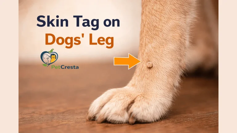
A small stable bump may be monitored after a vet exam. A growing, bleeding, crusting, or eye-touching lesion should be checked sooner. If the growth is on another body area, skin tag on dogs leg may be more relevant.
FAQ’s About Skin Tag on Dog Eye
A growth on a dog’s eyelid is any raised area, bump, flap, or small mass on the lid or lid margin. Some are benign skin tag like lesions, while others come from eyelid glands or nearby tissue. Because this area moves across the eye with every blink, even a small growth can matter more than it would on another part of the body.
Many skin tag like bumps near the eye are not dangerous in the sense of being aggressive disease. The concern is often mechanical rather than cancer related. A small lesion can still cause trouble if it rubs the cornea, creates tearing, changes blinking, or becomes inflamed over time.
Not every eyelid growth needs removal right away. A small stable bump that does not touch the eye may sometimes be monitored after a veterinary exam. Removal is more likely when the lesion gets larger, bleeds, crusts, changes eyelid shape, or causes redness, discharge, or rubbing.
A dog eyelid tumor may look like a small bump, a fleshy flap, a smooth nodule, a wart like growth, or a darker pigmented mass. Some remain narrow and raised. Others sit more broadly on the lid. Appearance can offer clues, but it cannot confirm the diagnosis without veterinary assessment and, in some cases, tissue review.
Most true skin tags and many eyelid growths do not simply disappear on their own. Some stay the same for a long time, while others slowly enlarge. A bump that changes, becomes irritated, or starts affecting the eye should not be watched casually without guidance.
Some small skin tags on other body areas may dry up, twist, or detach, but that is not something to expect or encourage near the eye. Eyelid growths should not be handled at home, even if they seem loose. A lesion that changes suddenly, bleeds, or partly tears away still needs a vet exam because the eyelid and cornea are easy to injure.
No. Cutting off a skin tag at home is not safe, especially near the eye. Home removal can cause bleeding, pain, eyelid damage, infection, and corneal injury. The safest approach is a veterinary exam to decide whether the bump can be monitored or whether professional removal is the better option.
Treatment depends on the type of growth and how it affects the eye. Some cases are monitored, while others are removed surgically to protect the eyelid margin and cornea. In selected cases, cryotherapy, laser treatment, or tissue sampling may also be part of the plan. Final diagnosis often depends on laboratory review of the tissue.
Cost varies with the size and location of the growth, the type of procedure, whether tissue testing is done, and the clinic or region. A simple removal is usually less involved than a case that needs more careful eyelid repair. The only reliable estimate comes after the eye and lesion have been examined in person.
Procedure time depends on the size and exact position of the growth. Small eyelid removals are often fairly quick, while more complex cases take longer if careful lid repair is needed. Recovery time also varies based on the procedure, the degree of irritation before surgery, and how well the dog avoids rubbing the eye afterward.
References
• American College of Veterinary Ophthalmologists, Canine Eyelid Masses
• PDSA, Eyelid Growths and Lumps on Dogs Eyelids
• Colorado State University Veterinary Teaching Hospital, Eyelid Mass